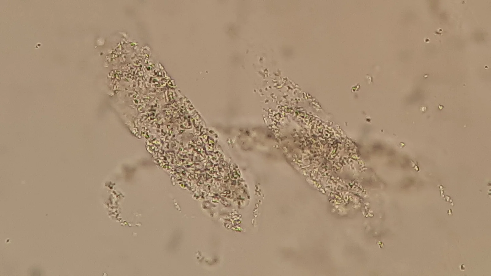
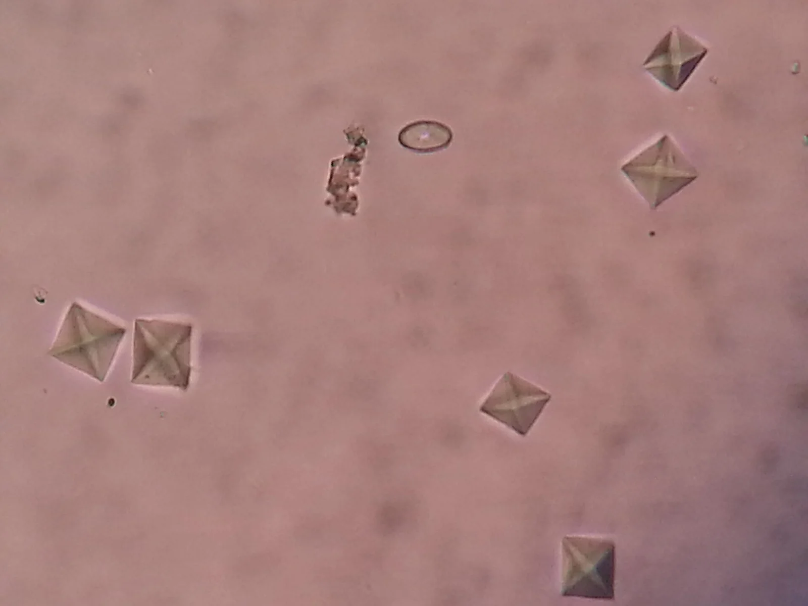
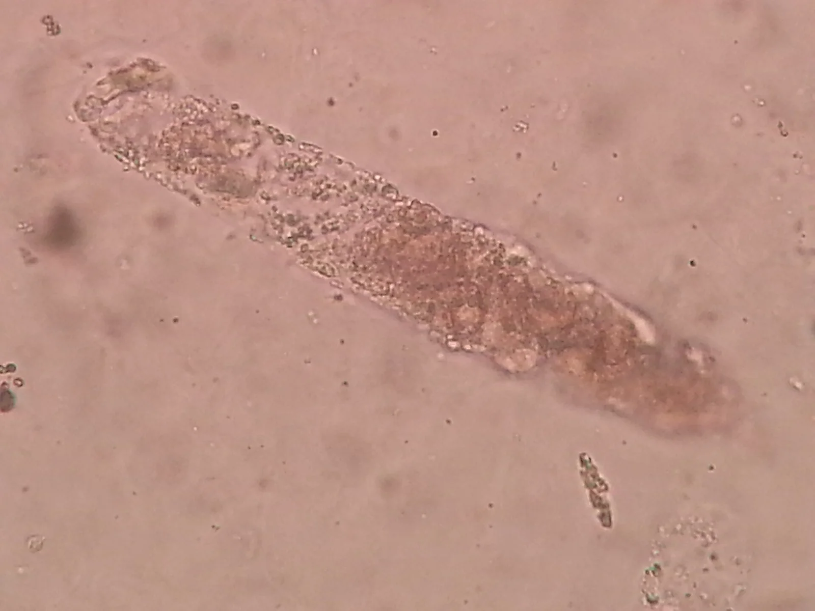
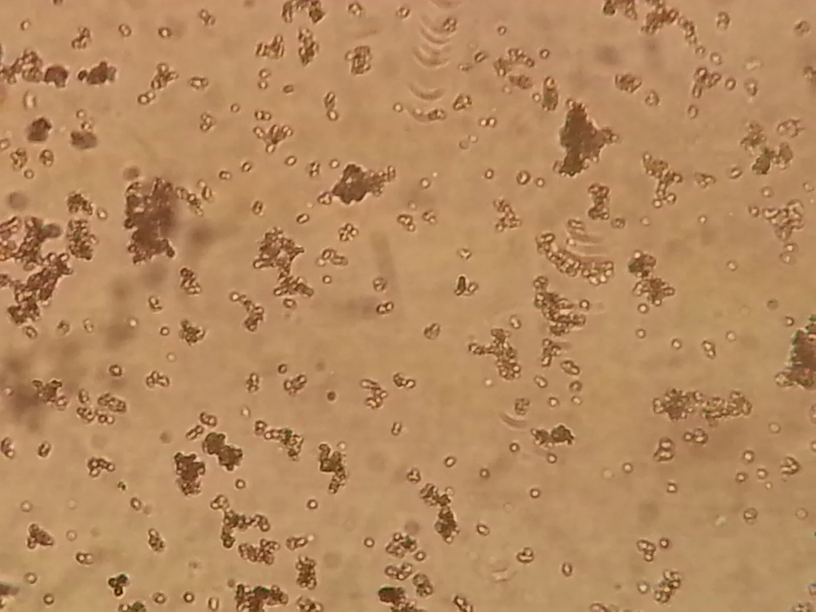
Muddy brown granular casts: the sediment of acute tubular necrosis. This field, in an oliguric patient with a rising creatinine, moves the diagnosis from prerenal to intrinsic.Ajay Kumar Chaurasiya, via Wikimedia Commons · CC BY 4.0 · sourceCalcium oxalate dihydrate: the envelope, a square with its diagonals drawn. Common in concentrated urine and in stone formers; in numbers after an ethylene glycol ingestion.Ajay Kumar Chaurasiya, via Wikimedia Commons · CC BY-SA 4.0 · sourceA coarse granular cast: a tubular mould packed with degenerated cell debris. One is a hint; many, pigmented, are acute tubular injury.Ajay Kumar Chaurasiya, via Wikimedia Commons · CC BY-SA 4.0 · sourceAmorphous urates: granular pinkish clumps in acid, concentrated or cooled urine. Usually a red herring, unless the story is tumour lysis.Ajay Kumar Chaurasiya, via Wikimedia Commons · CC BY-SA 4.0 · source
Hyperkalemia, rendered live
Peaked T waves with a widening QRS: the ECG nephrology cannot ignore.