The cheapest kidney biopsy in the building. Dipstick, sediment, and the urine chemistry that classifies AKI, sodium disorders, and acid-base problems.
The sediment, seen
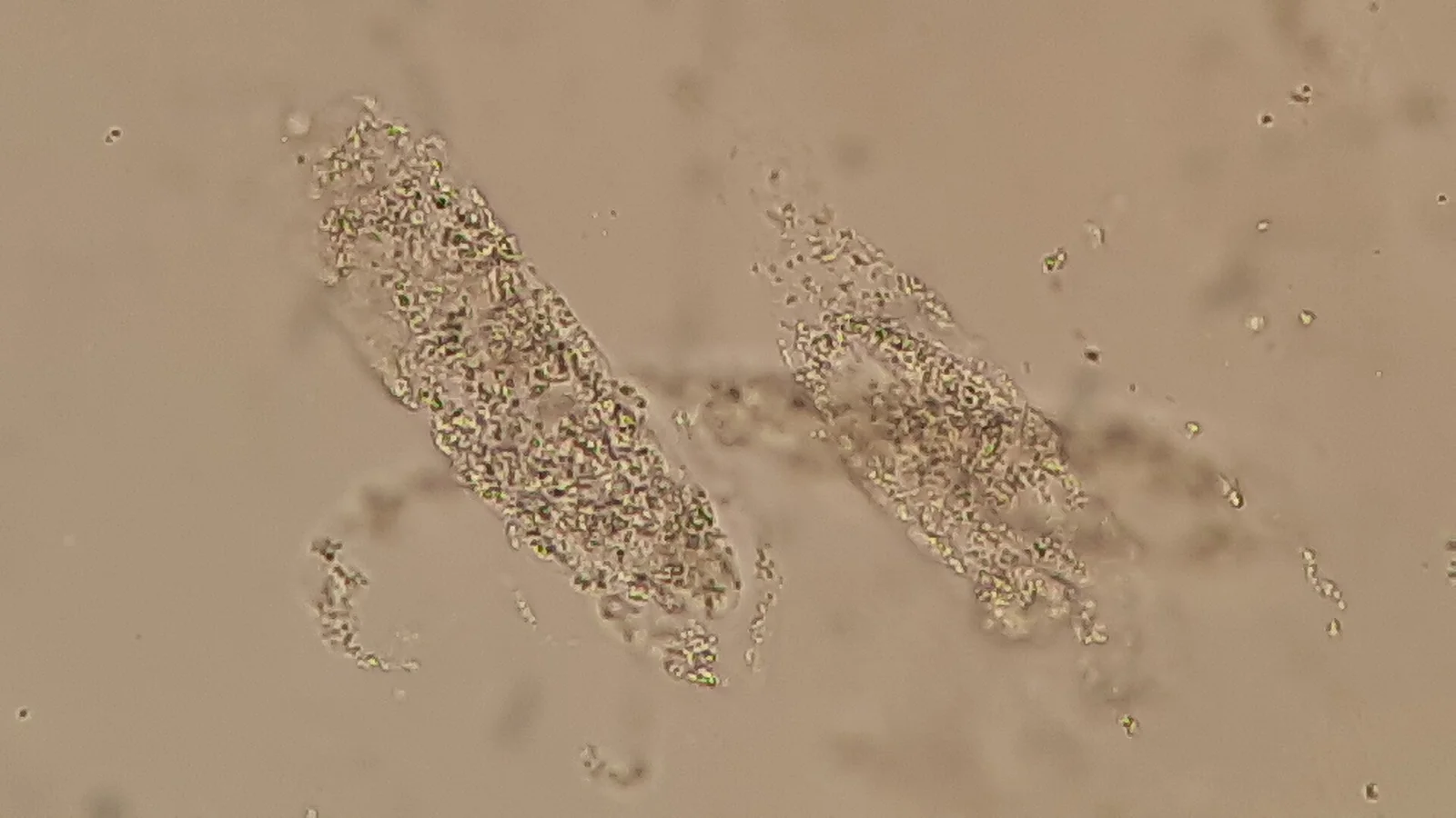
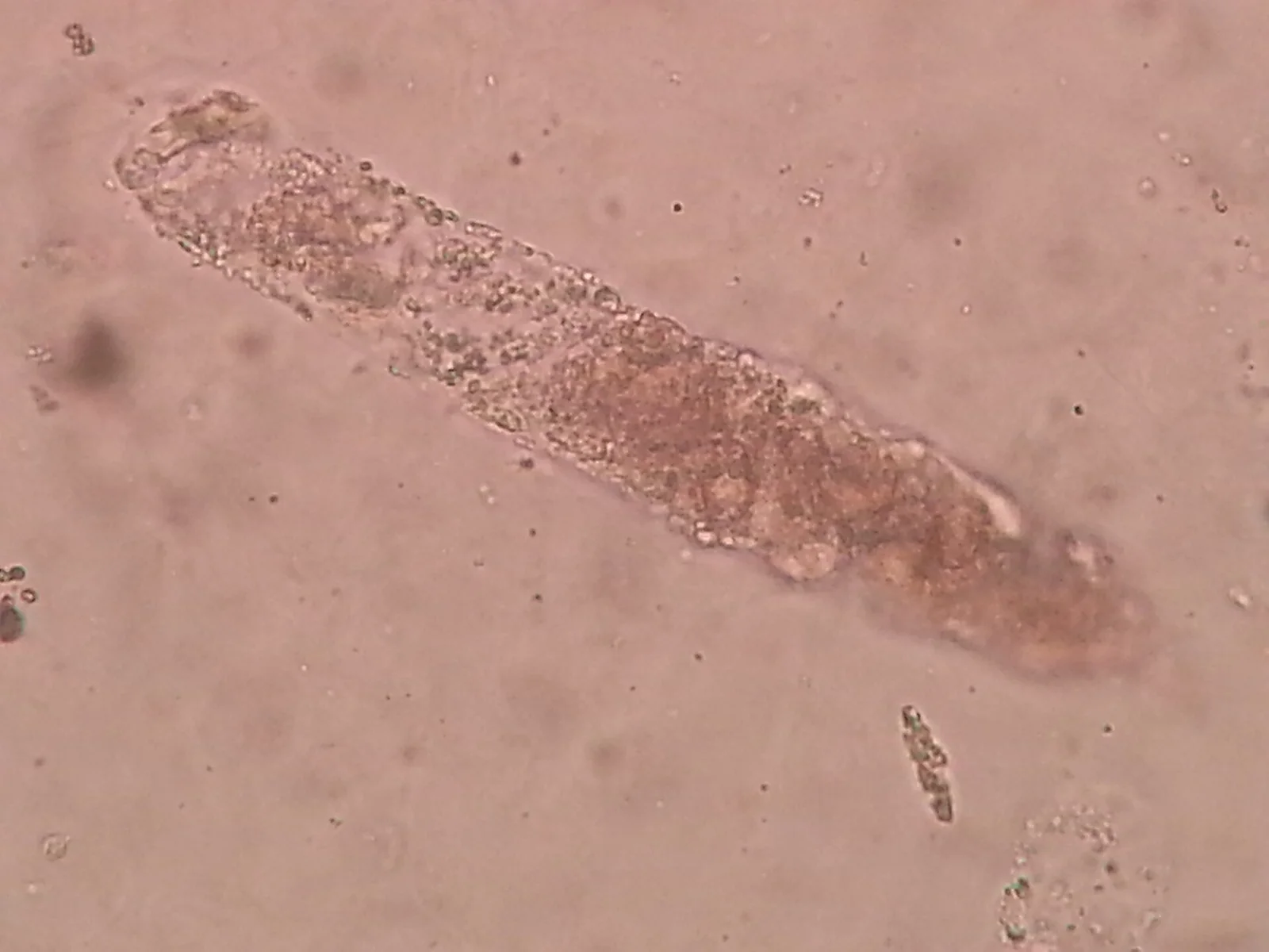
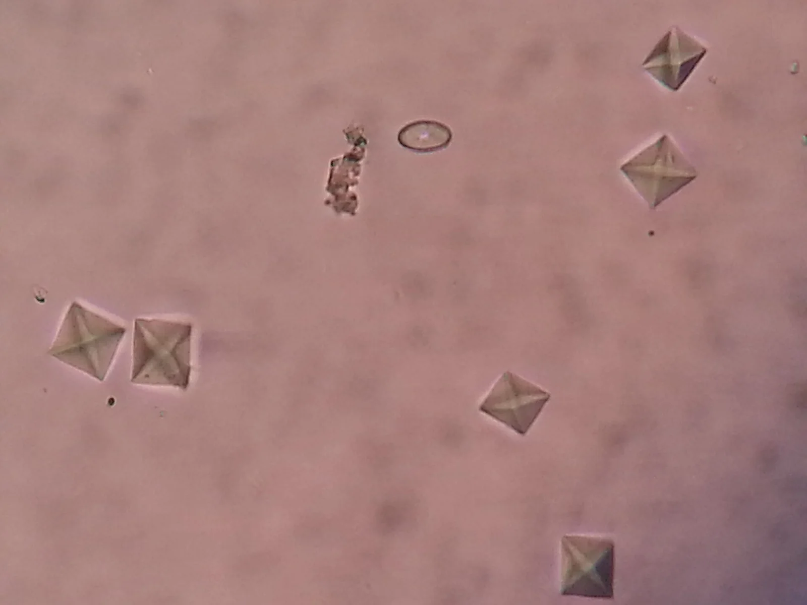
Casts and crystals under the microscope. The muddy brown granular cast is the one that separates tubular injury from a dry patient; the envelope crystal is calcium oxalate, the commonest stone former.
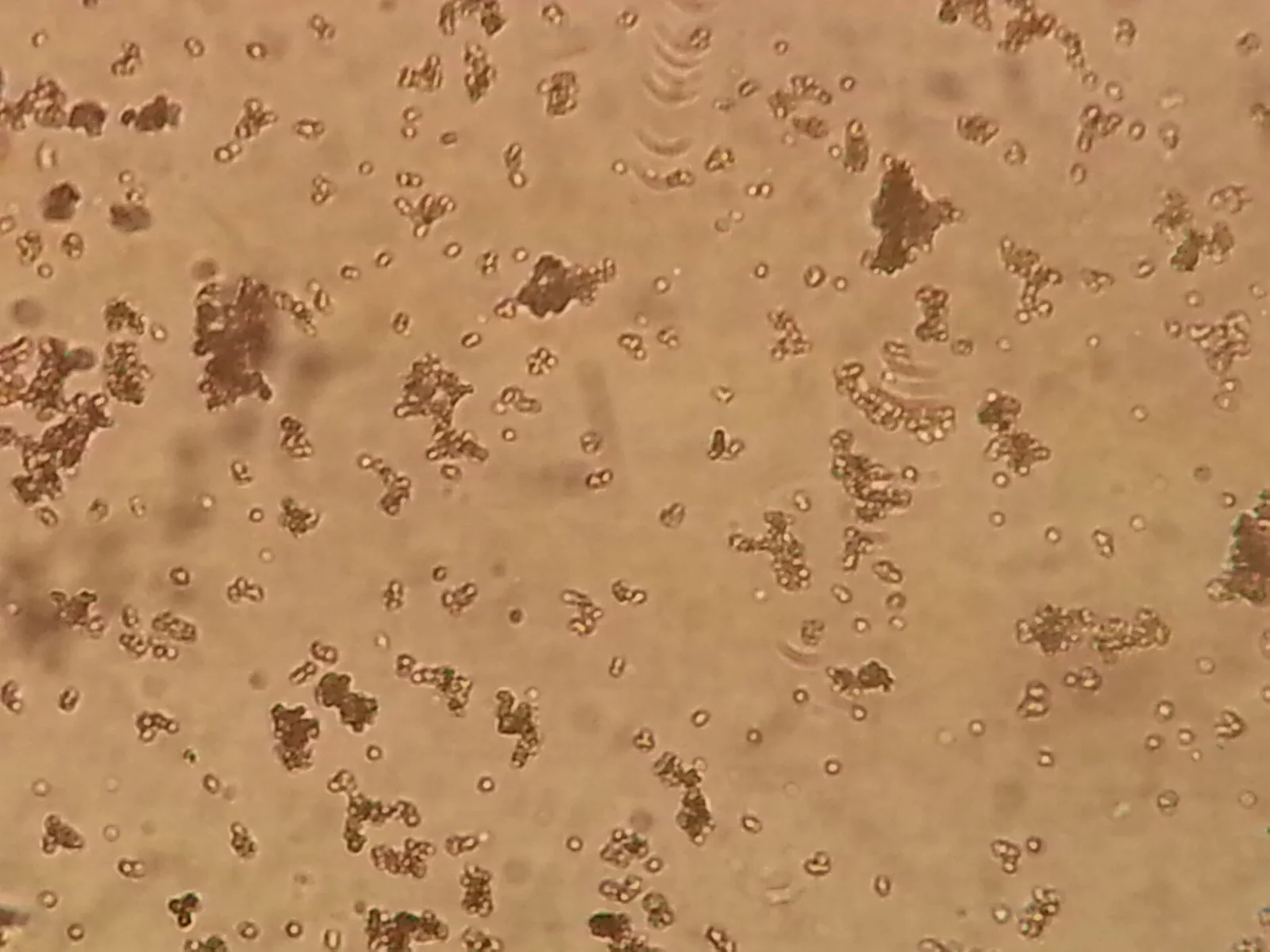
Muddy brown granular casts: the sediment of acute tubular necrosis. This field, in an oliguric patient with a rising creatinine, moves the diagnosis from prerenal to intrinsic.Ajay Kumar Chaurasiya, via Wikimedia Commons · CC BY 4.0 · sourceA coarse granular cast: a tubular mould packed with degenerated cell debris. One is a hint; many, pigmented, are acute tubular injury.Ajay Kumar Chaurasiya, via Wikimedia Commons · CC BY-SA 4.0 · sourceCalcium oxalate dihydrate: the envelope, a square with its diagonals drawn. Common in concentrated urine and in stone formers; in numbers after an ethylene glycol ingestion.Ajay Kumar Chaurasiya, via Wikimedia Commons · CC BY-SA 4.0 · sourceAmorphous urates: granular pinkish clumps in acid, concentrated or cooled urine. Usually a red herring, unless the story is tumour lysis.Ajay Kumar Chaurasiya, via Wikimedia Commons · CC BY-SA 4.0 · source
Dysuria and a positive dipstick
A 26-year-old with two days of dysuria and frequency, no fever or flank pain.
Dipstick
Leukocyte esterase
Positive
Nitrites
Positive
Blood
Trace
Protein
Negative
Glucose
Negative
Microscopy
Many white cells
Bacteria present
No casts
What does the nitrite result add beyond the leukocyte esterase?
AKI: what is the kidney trying to do?
The 74-year-old with pneumonia and a climbing creatinine from the lab module. You send urine studies before any fluid decision.
Dipstick
Protein
Trace
Blood
Negative
Microscopy
Bland sediment
Occasional hyaline casts
No granular casts
Urine chemistry
Urine sodium
12 mmol/L
Urine creatinine
9200 µmol/L
FENa
0.4 %
Urine osmolality
620 mOsm/kg
What is this kidney doing?
The urine that explains a sodium of 118
The euvolemic hyponatremic patient from the lab module. Her urine studies return.
Dipstick
Specific gravity
1.020
Urine chemistry
Urine osmolality
520 mOsm/kg
Urine sodium
64 mmol/L
Serum osmolality
246 mOsm/kg
Serum osmolality 246, urine osmolality 520. What does that pairing mean?
Normal gap acidosis: gut or kidney?
The traveler with diarrhea from the blood gas module. To prove the mechanism, you send spot urine electrolytes.
Dipstick
pH
5.2
Urine chemistry
Urine sodium
42 mmol/L
Urine potassium
28 mmol/L
Urine chloride
95 mmol/L
Urine anion gap
−25 mmol/L
The urine anion gap is negative. What does that say?
Tea-colored urine and puffy eyes
A 22-year-old presents with dark urine, facial swelling, and a BP of 168/98 two weeks after pharyngitis.
Dipstick
Blood
3+
Protein
2+
Leukocyte esterase
Negative
Nitrites
Negative
Microscopy
Dysmorphic red cells
Red cell casts
No crystals
Which single microscopy finding localizes the bleeding to the glomerulus?
The sediment after the arrest
Two days after a prolonged hypotensive episode in theatre, the creatinine is climbing and the urine output falling.
Dipstick
Blood
trace
Protein
1+
Leukocytes
negative
Nitrites
negative
Microscopy
Muddy brown granular casts, numerous
Renal tubular epithelial cells, occasional
No red cell casts, no dysmorphic red cells
Urine chemistry
Urine Na
68 mmol/L
FENa
2.8%
What does this sediment say about the mechanism of the AKI?
Dipstick blood, empty microscopy
The 79-year-old from the rhabdomyolysis lab case: dark urine after fourteen hours on the floor.
Dipstick
Blood
3+
Protein
1+
Leukocytes
negative
Nitrites
negative
Microscopy
No red cells seen
Pigmented granular casts
No red cell casts
How is a strongly heme-positive dipstick reconciled with a microscopy showing no red cells?
Frothy urine and swollen ankles
A 52-year-old with type 2 diabetes noticed frothy urine for months, and now ankle swelling to mid-shin. Albumin 24 g/L, creatinine 110.
Dipstick
Protein
4+
Blood
negative
Glucose
2+
Leukocytes
negative
Nitrites
negative
Microscopy
Oval fat bodies and fatty casts under polarized light ('Maltese crosses')
No red cell casts, no dysmorphic red cells
Heavy proteinuria, bland sediment, lipiduria, low albumin, edema. Which pattern is this, and what single quantification comes next?
Colic at four in the morning
A 38-year-old writhing with left loin-to-groin pain, vomiting, unable to lie still. Afebrile. Creatinine 96.
Which combination most raises the urgency in suspected ureteric colic?
The positive dipstick nobody was asking about
A routine dipstick on an 86-year-old with a long-term catheter, sent 'because it looked cloudy'. She is her usual self: comfortable, afebrile, eating breakfast.
Dipstick
Leukocytes
3+
Nitrites
positive
Blood
1+
Protein
trace
Microscopy
Numerous white cells and bacteria
No casts
Positive everything, asymptomatic patient, chronic catheter. What is the right move?