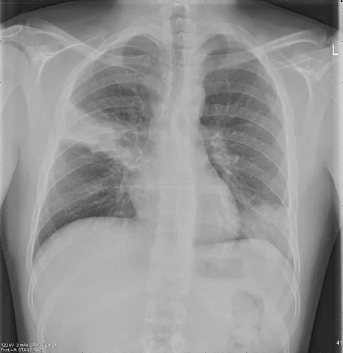
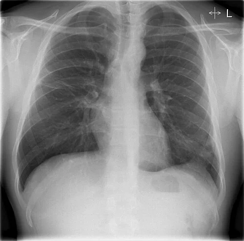
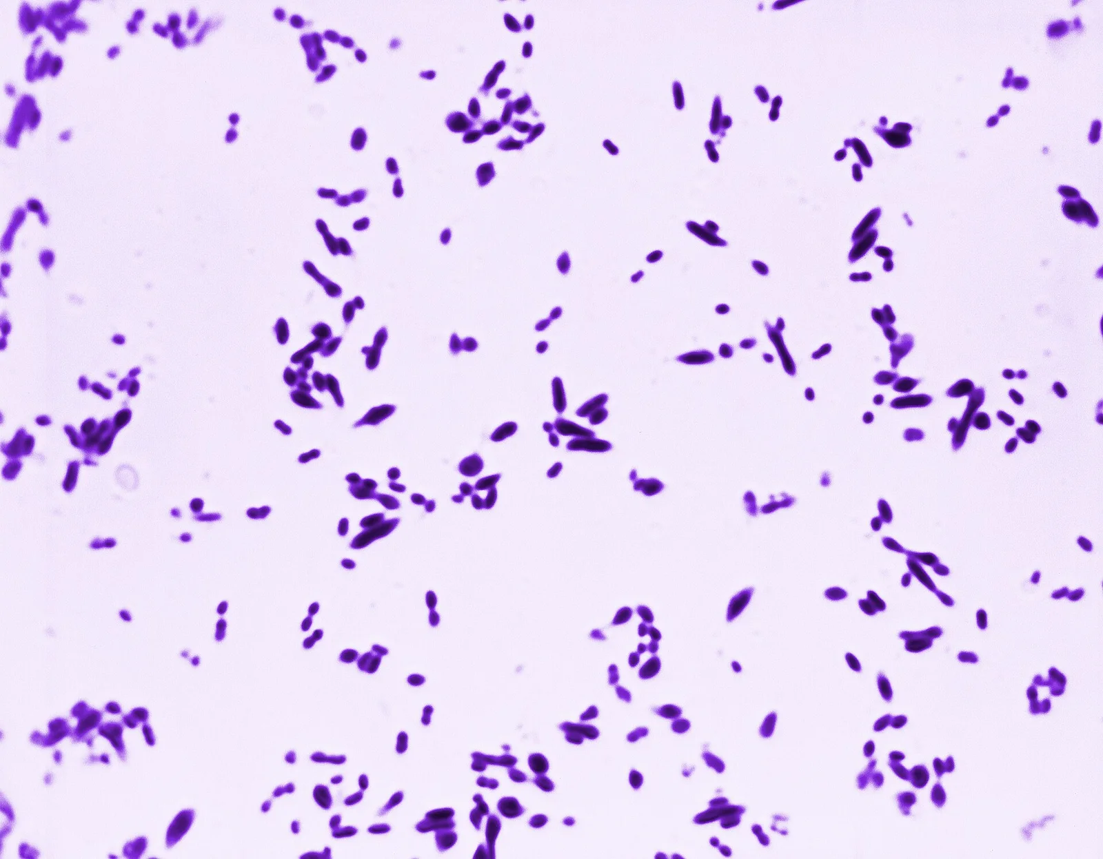
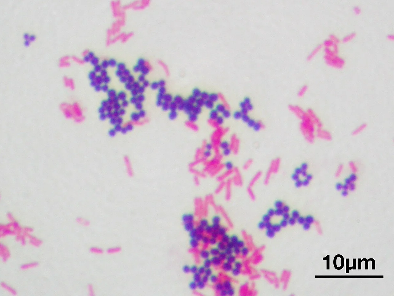
Community pneumonia at presentation: dense consolidation with air bronchograms. The film to keep, because the follow-up only means something beside it.Jtechr, via Wikimedia Commons · CC BY-SA 3.0 · sourceThe same patient after antibiotics: the consolidation has largely cleared. Radiographic resolution lags clinical recovery by weeks, which is why the follow-up film waits.Jtechr, via Wikimedia Commons · CC BY-SA 3.0 · sourceGram positive diplococci, lancet shaped, in pairs: Streptococcus pneumoniae. In cerebrospinal fluid this picture names the organism hours before the culture does.Arnold Kaufman, via Wikimedia Commons · CC0 · sourceBoth answers on one slide: gram positive cocci in clusters (purple) and gram negative rods (pink). The colour is the cell wall; the shape and grouping are the organism.Y tambe, via Wikimedia Commons · CC BY-SA 3.0 · source