Presentations
Approach to fever in the hospital
The same eight steps, every presentation: threats first, then the history and exam that discriminate, one honest sentence, a mechanism-grouped differential, and the tests read in order.
Immediate threats
- Septic shock physiology
- Neutropenic fever: antibiotics within the hour
- Meningitis; necrotizing soft tissue infection
- The non-infectious killers wearing fever: PE, drug fever syndromes
History that discriminates
- Localizing symptoms walked head to toe
- Lines, catheters, devices, recent procedures: each one a suspect
- Immunosuppression inventory; travel and exposures
- New drugs (drug fever, serotonin and neuroleptic syndromes)
Examination
- Every line site and every drain, uncovered and looked at
- Lungs, abdomen, skin folds, joints, calves
- The back: sacral wounds hide from busy teams
Problem representation
New fever on ward day 3 in a stented, recently instrumented urinary system (urosepsis weight, source-control question) versus fever with a new murmur and split hands (endocarditis weight).
Differential, by mechanism
Infectious
- Urinary (catheter)
- Pneumonia (aspiration weight in the frail)
- Line infection
- Surgical site and collections
- C. difficile
- Endocarditis
Non-infectious
- VTE
- Drug fever
- Gout flare
- Transfusion reactions
- Malignancy and connective tissue disease
Investigations
- Cultures before antibiotics: blood times two, urine, and anything drainable
- Directed imaging over reflex pan-scanning, but a low threshold in the deteriorating
- Lactate as the severity meter
Interpretation
- Fever plus hypotension runs the sepsis clock regardless of the eventual source
- A fever without localization on day one localizes on day two: re-examine
Next steps
- Empiric coverage matched to the suspected source and local patterns, de-escalated on data
- Source control is asked about early and out loud
What the images look like
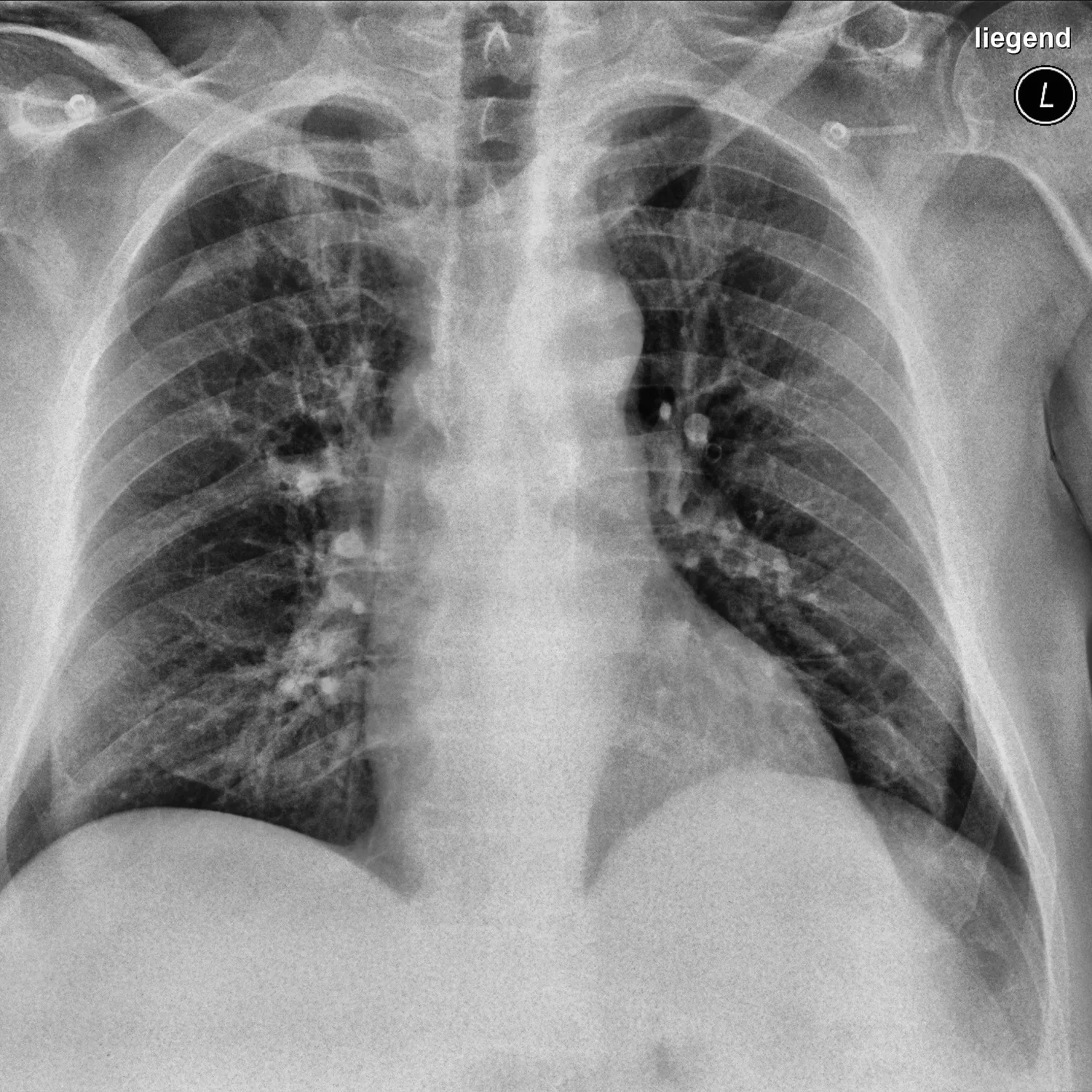
Connected
Take it further
Copy this page as study notes, then paste them into QSpace for practice questions or AnkiGen for flashcards.