Pediatric presentation

Cough and respiratory distress in children

Bronchiolitis, croup, asthma and pneumonia are separated by the age, the sound and the effort, not by the X-ray.

  1. What are you worried about?

    • Impending respiratory failure: exhaustion, a quiet chest, drowsiness, a falling respiratory rate
    • Upper airway obstruction: stridor at rest, drooling, tripod position
    • Apnea in the young infant with bronchiolitis or pertussis
    • The inhaled foreign body
  2. Differential, by age

    Newborn birth to 28 days

    1. Sepsis presenting as breathing difficulty
    2. Congenital heart disease
    3. Bronchiolitis with apnea
    4. Pertussis

    Infant 1 to 12 months

    1. Bronchiolitis
    2. Pneumonia
    3. Pertussis
    4. Heart failure from a shunt lesion

    Toddler 1 to 3 years

    1. Croup
    2. Viral-induced wheeze
    3. Foreign body aspiration
    4. Pneumonia

    Preschool 3 to 5 years

    1. Asthma
    2. Croup
    3. Pneumonia
    4. Foreign body

    School age 6 to 11 years

    1. Asthma
    2. Pneumonia, including Mycoplasma
    3. Anaphylaxis

    Adolescent 12 to 17 years

    1. Asthma
    2. Pneumonia
    3. Pneumothorax
    4. Vaping-associated lung injury
    5. Pulmonary embolism with risk factors
  3. Key history

    • Onset and tempo: sudden (foreign body, anaphylaxis, pneumothorax) versus days of coryza first (bronchiolitis, croup)
    • The sound: bark, stridor, wheeze, grunt, whoop
    • Feeding: the volume taken, coming off the breast or bottle to breathe
    • Fever, colour changes, apneas, the night pattern (croup peaks at night)
    • Prematurity, chronic lung disease, congenital heart disease, previous wheeze, asthma control, smoke exposure
    • Immunizations (pertussis, Haemophilus influenzae type b, pneumococcus)
  4. Key examination

    • Work of breathing from the doorway: rate, nasal flaring, head bobbing, tracheal tug, retractions (subcostal, intercostal, suprasternal), grunting
    • Ability to feed, talk, or play; level of alertness
    • Where the noise lives: inspiratory (upper airway) versus expiratory (lower airway)
    • Air entry and its symmetry; crackles; the silent chest
    • Saturation, and how much oxygen it is taking to hold it
    • Hydration and perfusion
  5. Red flags

    • Saturation below 90 percent, or a rising oxygen requirement
    • Grunting, head bobbing, severe retractions, a silent chest
    • Apnea or cyanotic episodes, especially under 3 months or ex-preterm
    • Exhaustion: drowsiness, a slowing respiratory rate, rising carbon dioxide
    • Stridor at rest, drooling, inability to swallow
    • Intake below about half of usual, or no wet diaper in 12 hours
  6. Initial investigations

    • Bronchiolitis and croup are clinical diagnoses: no X-ray, no viral swab, no blood test routinely
    • Chest X-ray for focal signs, a suspected foreign body, a first severe wheeze, or failure to improve
    • A blood gas when exhaustion is a question
    • Inflammatory markers and cultures only when bacterial pneumonia or sepsis is suspected
  7. How to interpret them

    • Wheeze responsiveness to bronchodilator separates asthma physiology from bronchiolitis, which does not respond
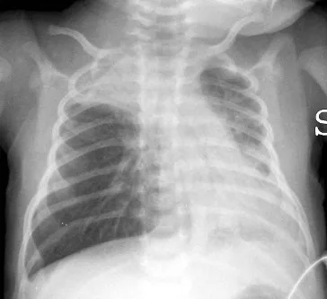
    • Hyperinflation on a bronchiolitis film is expected and is not pneumonia
    • A normal saturation on high-flow oxygen is not a normal child
  8. When the child is deteriorating

    • Escalate oxygen delivery stepwise; high-flow nasal cannula in bronchiolitis; nebulized epinephrine and dexamethasone in croup
    • Asthma: bronchodilator, steroid, then magnesium and senior review; a falling heart rate with a quiet chest is peri-arrest
    • Know the numbers that trigger the call: oxygen requirement, respiratory rate trend, mental status, gas

See it

Frontal chest radiograph of an infant with bronchiolitis showing hyperinflated lungs, flattened diaphragms and patchy atelectasis
Bronchiolitis in a 16-day-old: hyperinflation with flattened diaphragms and bilateral atelectasis (right apical, left basal). The film is not needed to diagnose bronchiolitis; it is here to show what hyperinflation looks like in an infant.Di Nardo M, Perrotta D, Stoppa F, Cecchetti C, Marano M, Pirozzi N (Journal of Medical Case Reports, via Wikimedia Commons) · CC BY 2.0 · source

Watch

Recognizing Respiratory Distress by M. Kleinman

OPENPediatrics (Boston Children's Hospital) · embedded with attribution; their teaching, not ours

The clinical signs of respiratory distress in a child, from the effort to the sounds to the level of consciousness, taught by a pediatric intensivist.

Watch for

  • The order in which effort, sounds and mental status are read
  • What separates distress from failure
  • Why the respiratory rate alone misleads
Watch on YouTube

Module 3: Retractions

Stanford Center for Health Education · embedded with attribution; their teaching, not ours

Chest indrawing in a child, shown and named: subcostal, intercostal, suprasternal, and the flaring and head bobbing that go with it.

Watch for

  • Where each retraction shows
  • Head bobbing and nasal flaring in an infant
  • How the picture changes as the child tires
Watch on YouTube

Conditions under this presentation

Pediatric chest films: hyperinflation, consolidation, the foreign body

The adult frame, for contrast