The same eight steps, every presentation: threats first, then the history and exam that discriminate, one honest sentence, a mechanism-grouped differential, and the tests read in order.
01
Immediate threats
Hypoxia needing oxygen now (SpO2, work of breathing, mentation)
Air entry, wheeze, crackles, dullness (the pulmonary column)
Calves, and conjunctivae for pallor
04
Problem representation
Acute pleuritic dyspnea with hypoxia and a clear chest in a post-operative patient (points at PE) versus subacute orthopneic dyspnea with a raised JVP and crackles (points at failure).
05
Differential, by mechanism
Cardiac
Heart failure
ACS
Arrhythmia
Tamponade
Valve disease
Pulmonary
Pneumonia
COPD/asthma exacerbation
PE
Pneumothorax
Effusion
ILD
Hematologic / metabolic
Anemia
Acidosis with compensatory tachypnea (DKA, sepsis)
Other
Anxiety (diagnosis of exclusion)
Neuromuscular weakness
Upper airway obstruction
06
Investigations
SpO2 and blood gas when saturation or mentation is off
ECG and troponin where cardiac weight exists
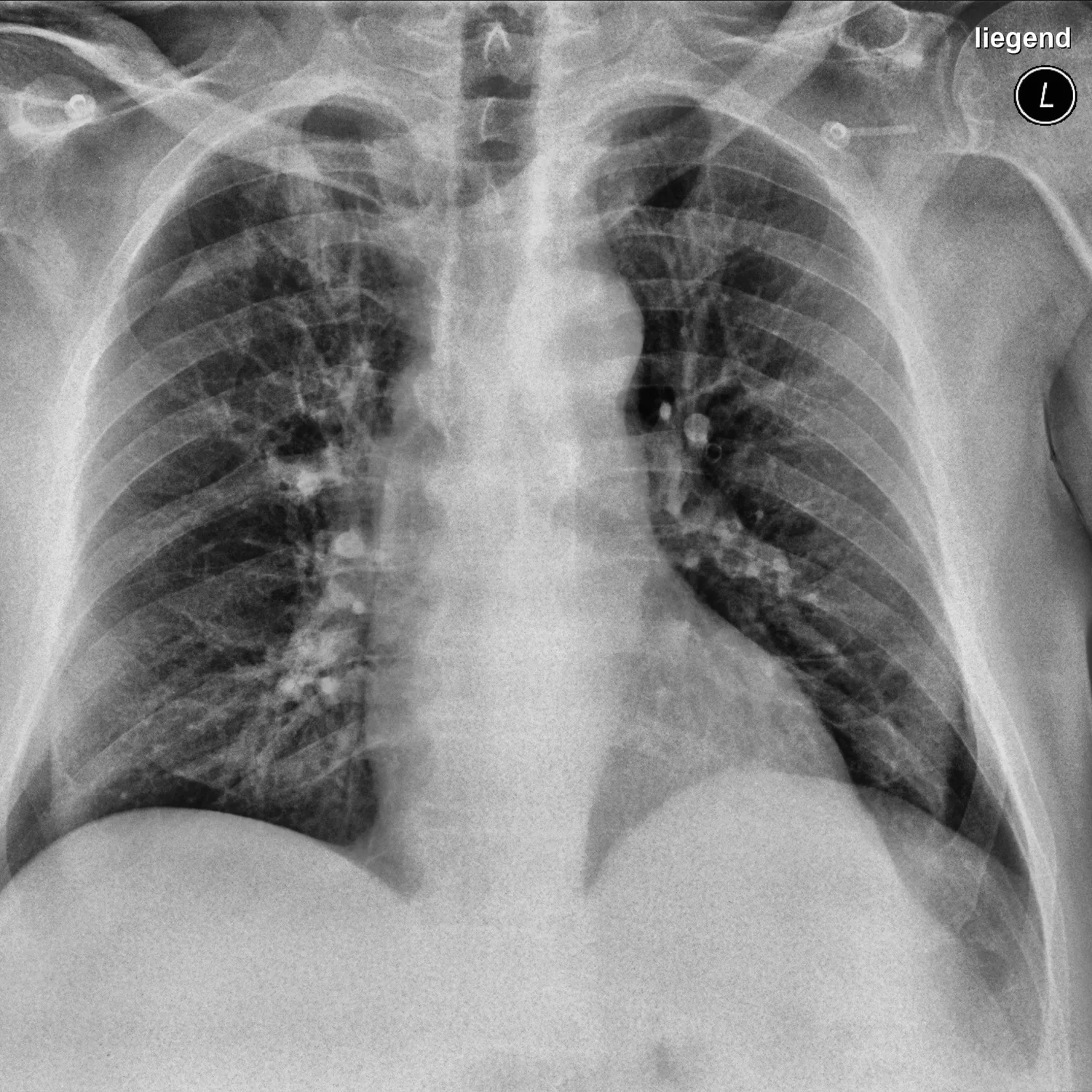
CXR: the single highest-yield image
NT-proBNP when failure versus lung is genuinely uncertain
Wells-gated D-dimer or CTPA for PE weight
CBC for anemia; PFTs for the chronic outpatient course
07
Interpretation
Hypoxia with a clear exam and film pushes toward PE or early shunt physiology
The gas splits ventilation failure (high PaCO2) from oxygenation failure (wide A-a gradient)
The trend of work of breathing outranks any single number
08
Next steps
Treat the threat first (oxygen, then the specific lesion)
Every dyspnea gets a named leading diagnosis and a stated reassessment time
What the images look like
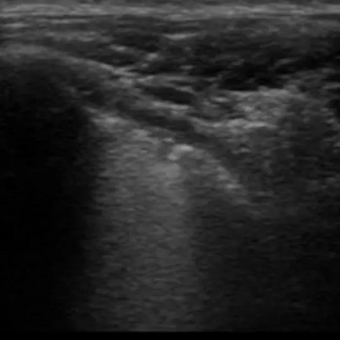
Chest radiograph in COVID-19 viral pneumonia: patchy bilateral opacities.Hannover Medical School COVID-19 Image Repository (via the ieee8023 covid-chestxray-dataset) · CC BY 3.0 · sourceViral pneumonia on lung ultrasound: irregular pleura with confluent vertical artifacts.COVID-19 Lung Ultrasound Dataset (Born et al.) · CC BY 4.0 · source