ECG library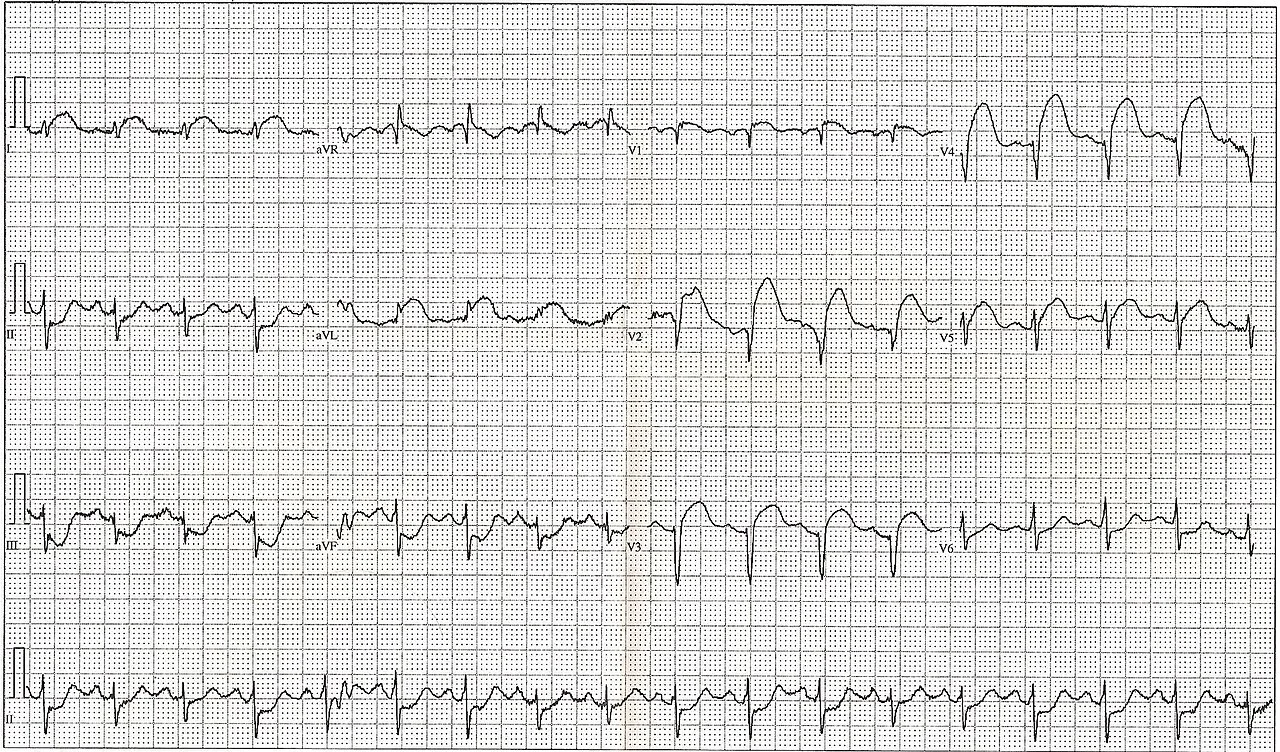
Displaced, via Wikimedia Commons · Public domain · source
Anterior ST-elevation myocardial infarction
coreA 54-year-old smoker, 40 minutes of chest pain, pale and sweating.
25 mm/s · 10 mm/mV · synthetic tracing
100%
Calipers:2000 ms50.0 small boxes30 bpm if R-RDrag the orange markers. One small box = 40 ms.
Recorded tracingA real 12-lead of this rhythm. The strip above is a drawn model; this is the paper.
Key findings
- STE V1 to V4
- Evolving septal Qs
- The LAD supplies the most myocardium
Learning points
- Territory maps to artery: septal-anterior V1 to V4 is the LAD's signature.
- Q waves forming during pain mean myocardium is already dying; the clock is not theoretical.
Connected